Structure and mechanogating of the mammalian tactile channel PIEZO1
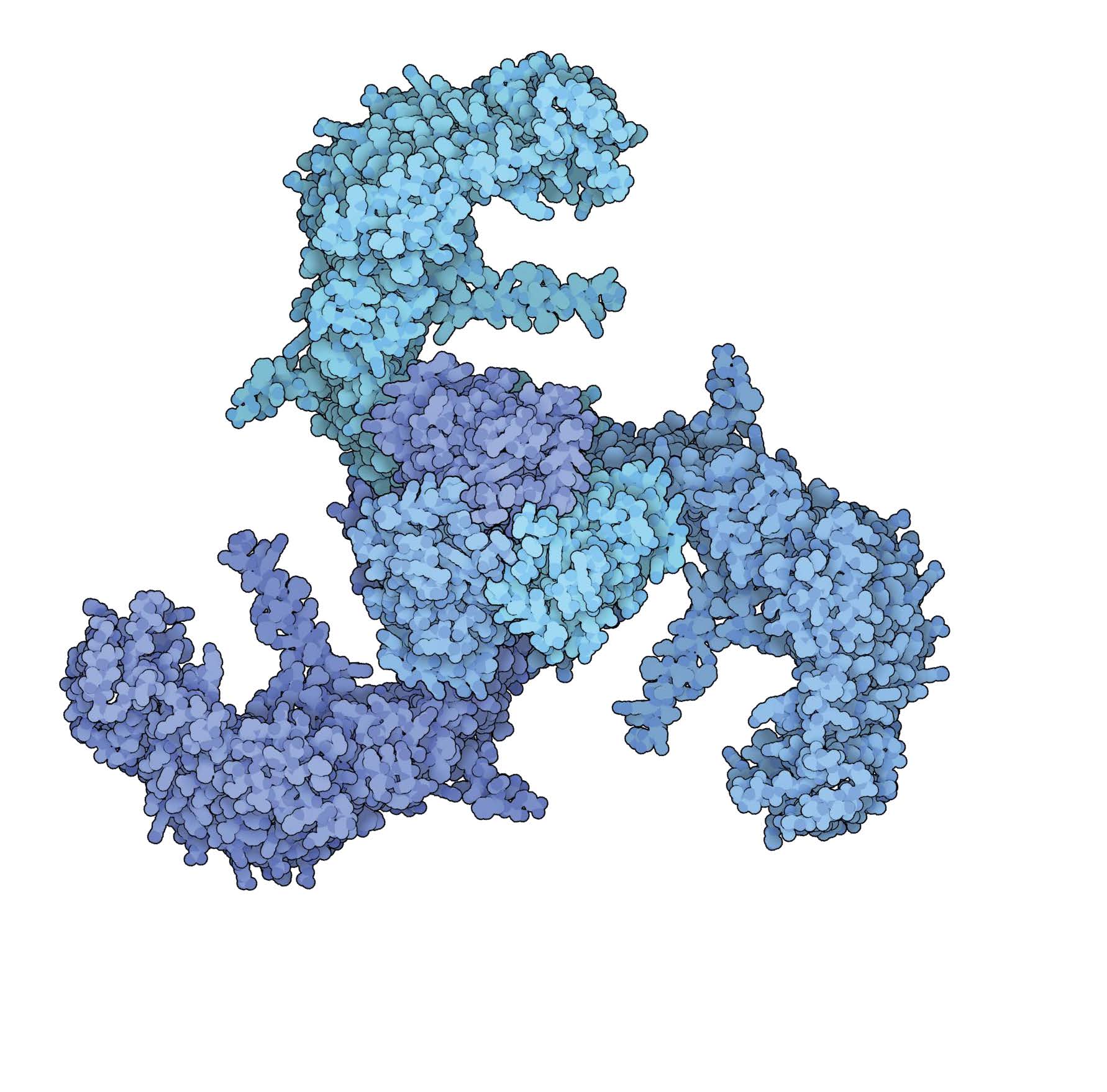
In some animals, three blade-like proteins together form the Piezo1 channel, which helps their cells to sense touch. Credit: David S. Goodsell/RCSB PDB.
Structure and mechanogating of the mammalian tactile channel PIEZO2
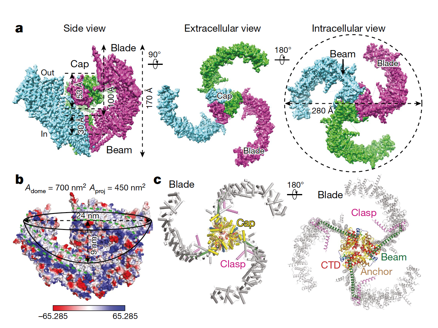
The homotrimeric structure of PIEZO2. a, The indicated views of the sharpened map (2.78σ contour level) with a resolution of 3.6–3.8 Å. b, A side view of the surface electrostatic potential, showing the hydrophobic transmembrane region (marked by green dashed lines). The mid-plane opening diameter, depth, surface area (Adome) and projection area (Aproj) of the illustrated dome are labelled. The colour bar indicates the surface electrostatic potential, ranging from negative (red) to positive (blue). c, Cartoon models with the major structural domains coloured and labelled.
The structural coordinates of mouse PIEZO2 have been deposited in the PDB under the accession 6KG7. The cryo-EM map was deposited into the Electron Microscopy Data Bank (EMDB) under the accession 9975.
Wang, L., Zhou, H., Zhang, M. et al. Structure and mechanogating of the mammalian tactile channel PIEZO2. Nature 573, 225–229 (2019). https://doi.org/10.1038/s41586-019-1505-8